
Magnetic resonance imaging is an important examination modality for the prevention, diagnosis and aftercare of tumors. Thanks to this method, not only benign changes but also malignant diseases can be detected and treated at an early stage. In my radiology practice, I use imaging to clarify oncological issues.
Cancer can have many causes. Pre-existing conditions, genetic predisposition, lifestyle and environmental influences can all play a role in its origin. Cancerous disease is characterised by the proliferation of malignant tissue. This means that cells can grow uncontrollably and displace or destroy the surrounding healthy tissue. There is also the risk of cells spreading in the body. This is referred to as metastases and, in the case of certain entities (e.g. lymphoma) as manifestations.
One of the most important tasks of imaging is tumour diagnosis – and in the best case the early detection of signs of malignant disease. In the case of cancerous disease, oncological imaging with tumour and propagation diagnostics (staging) provides an optimal basis for individualised therapy planning and, if necessary, monitoring of the therapy.

Oncological imaging
Cancer diagnostics using MRI
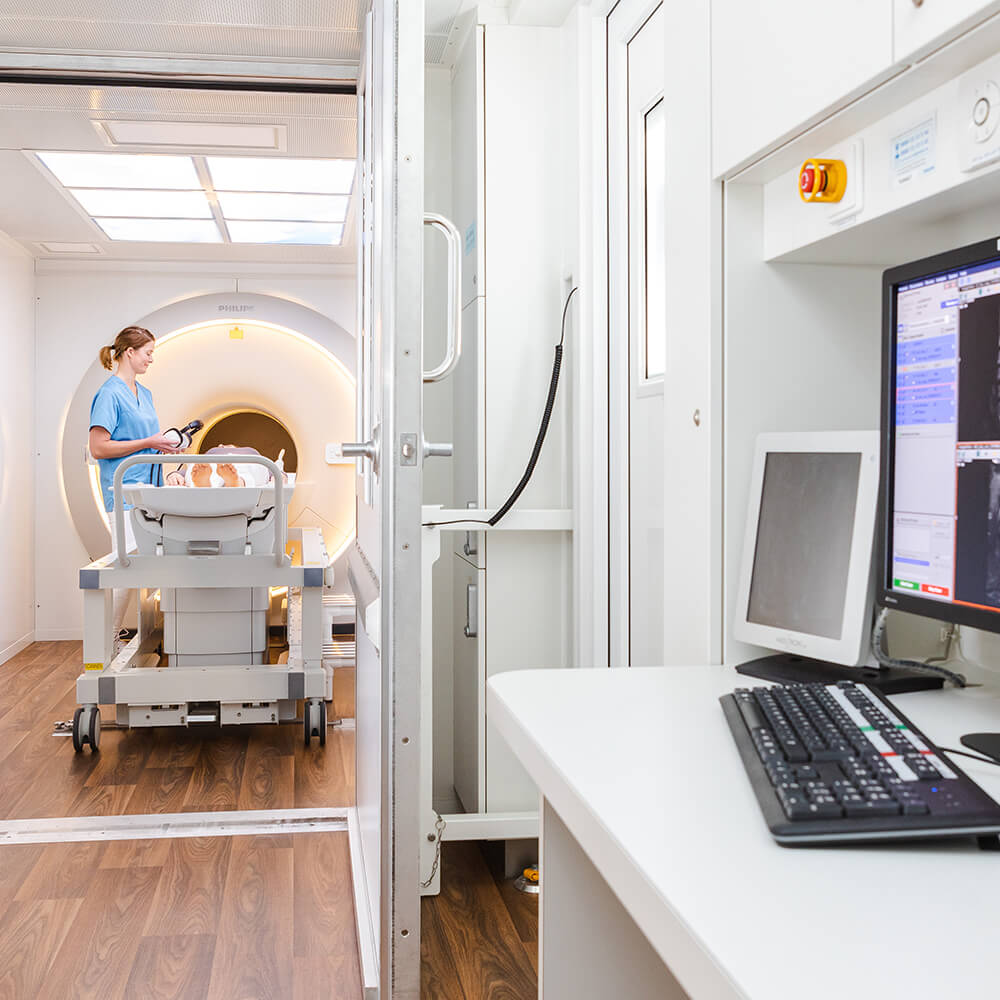
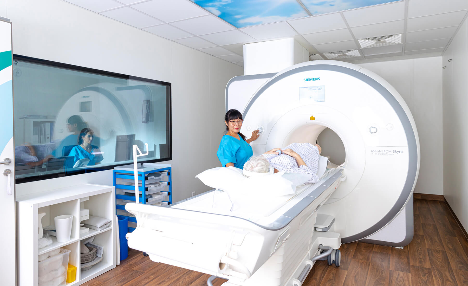
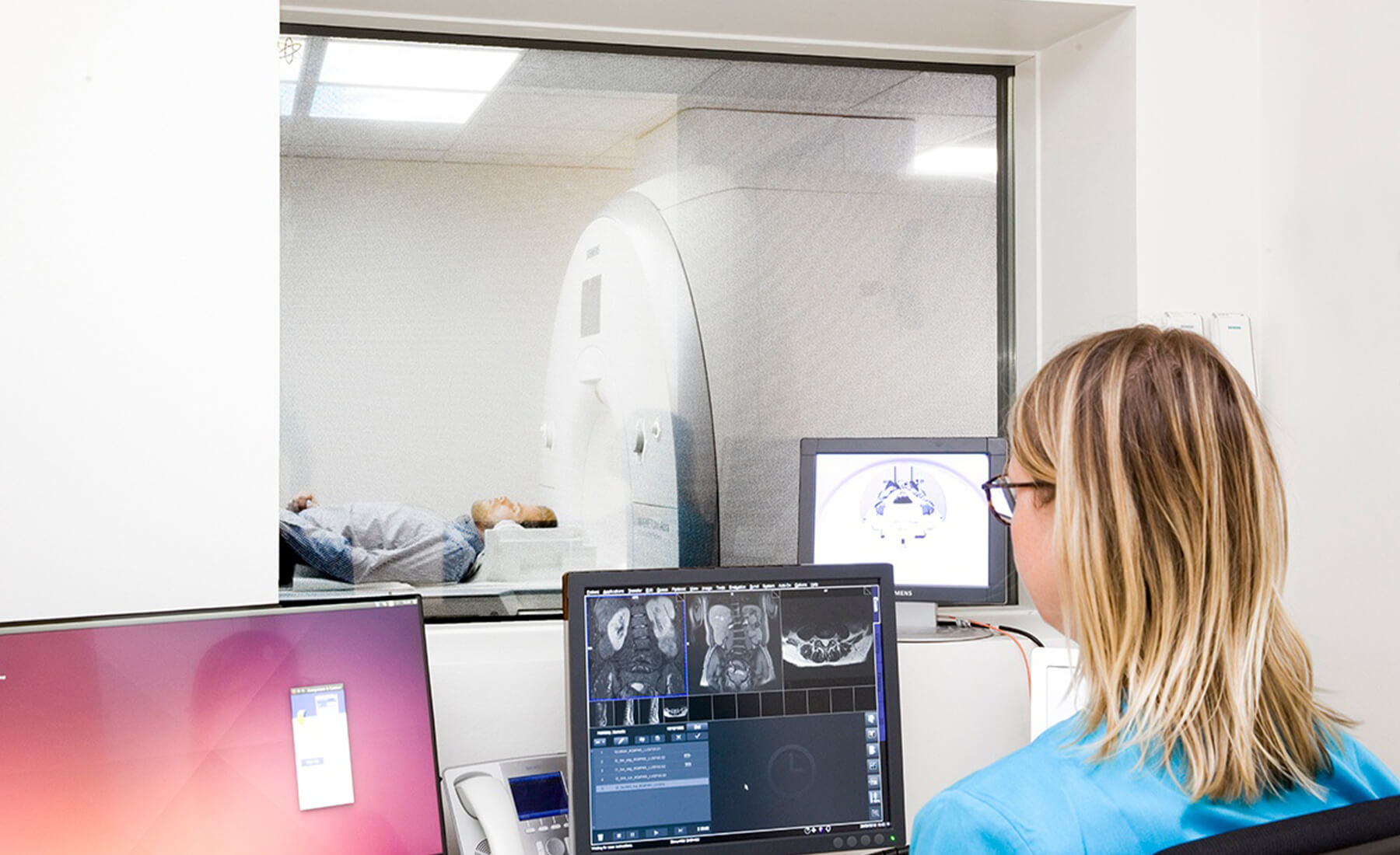
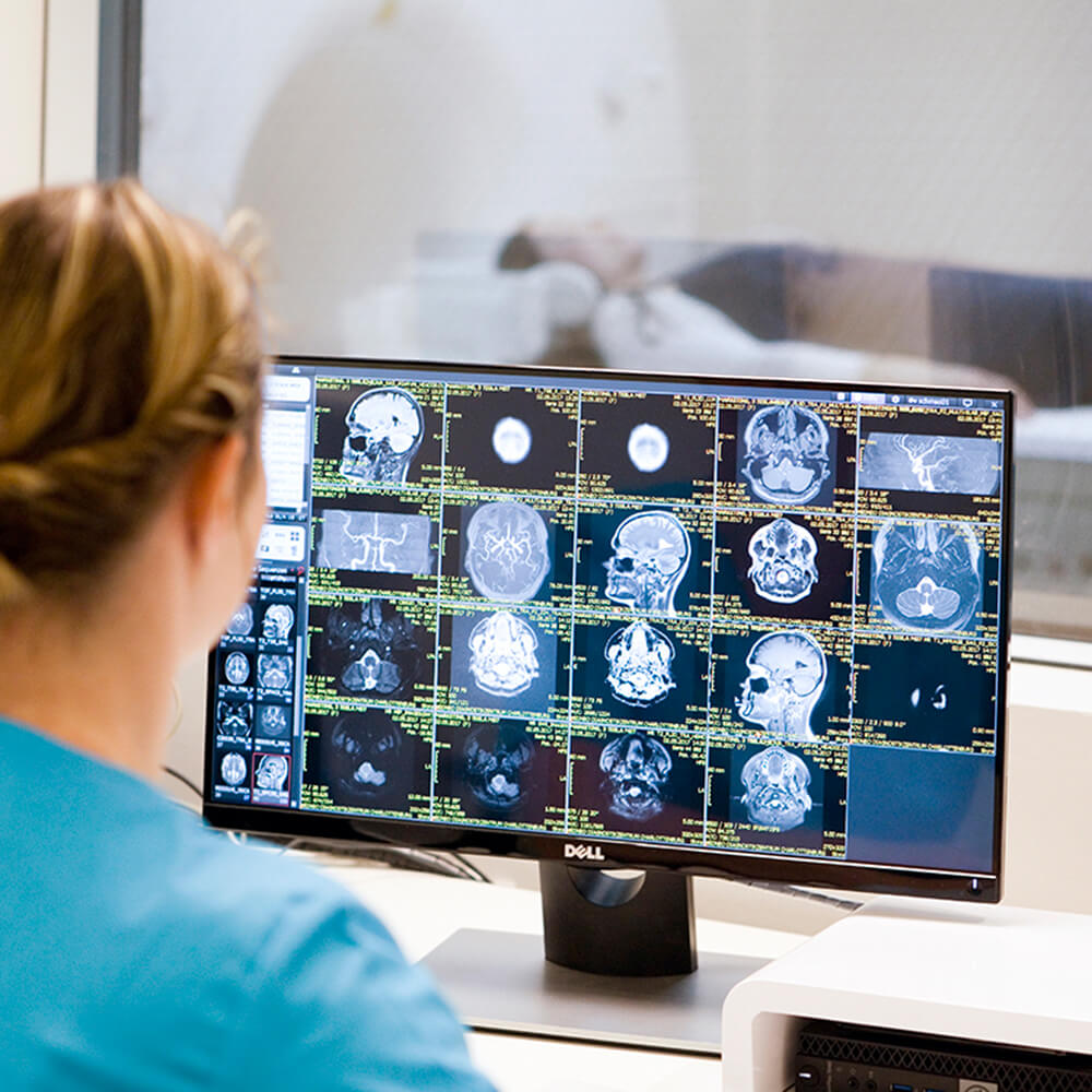
Oncological imaging by MRI
Magnetic resonance imaging is also important for cancer diagnostics. Many questions in oncology can be specifically clarified with special MRI diagnostics.
The soft tissues, the organs and the bone marrow are depicted in a highly differentiated way without radiation exposure. Tumorous changes in the brain and spinal cord, breast cancer, prostate cancer, small tumours in the liver and pancreas can be detected very well, for example. A strength of MRI here is usually also the relevant ability to assess tumour boundaries. MRI is the preferred imaging method for spinal cord compression.
Oncological imaging by CT
Computed tomography allows me to very quickly and reliably detect whether there is a tumour in the region of the body under examination or whether there is an indication of spreading. In this case, large or multiple body regions can also be comprehensively depicted in a single examination.
CT is particularly suitable for evaluating tumours in the lungs and abdomen. It offers high specificity in detecting bone metastases. Bone metastases most frequently occur in renal cell, bronchial, breast or prostate carcinoma. CT allows stability assessment in cases of extensive metastatic bone destruction and is also useful in biopsy confirmation of bone lesions.
Imaging important for cancer aftercare
Aftercare for long-term health
Once cancer has been detected and successfully treated, follow-up care is essential to detect any recurrence as early as possible.
In addition to physical examinations and laboratory tests, imaging is an essential component in the aftercare concept. The method of choice with specific planning of the examination is oriented on the tumour entity and the individual situation of the patient.